Home
Uncategories
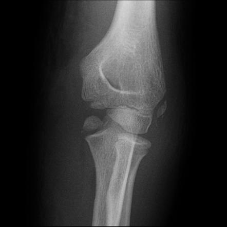
Medial Humeral Epicondyle Fracture : Medial Epicondyle Nonunions In Children Case Report With Overview And Management Abstract Europe Pmc / Internal oblique view to evaluate displacement.
Medial Humeral Epicondyle Fracture : Medial Epicondyle Nonunions In Children Case Report With Overview And Management Abstract Europe Pmc / Internal oblique view to evaluate displacement.
Medial Humeral Epicondyle Fracture : Medial Epicondyle Nonunions In Children Case Report With Overview And Management Abstract Europe Pmc / Internal oblique view to evaluate displacement.. Flex your wrist toward your forearm. The treatment of isolated, displaced fractures of the medial humeral epicondyle in children is controversial. Point tenderness over or just distal to the medial humeral epicondyle. In a large combined series of 5228 fractures of the distal humerus, medial epicondyle fractures constituted 14.1% of all distal humeral fractures and 11.5% of all fractures occurring around the elbow. Related online courses on physioplus.
Distal humerus fracture is very common in pediatric patients; Introduction to telehealth and paediatrics online course: Operative management of the pediatric medial epicondyle fracture. The medial and lateral epicondyles are easily palpable, and form the sites of origin for the forearm flexors of the anterior compartment and forearm. Medial epicondyle fractures are much more common than medial condyle fractures.
Medial Epicondyle Fracture Radiology Reference Article Radiopaedia Org from prod-images-static.radiopaedia.org The treatment of isolated, displaced fractures of the medial humeral epicondyle in children is controversial. Treatment of pediatric humeral medial epicondyle fractures is controversial. A fracture of the medial epicondyle of the elbow that is the third most common fracture seen in children and is usually seen in boys between the age of 9 and 14. Kilfoyle classified these fractures into three types according to the degree of displacement 5. The medial epicondyle of the humerus is an epicondyle of the humerus bone of the upper arm in humans. The management of displaced medial humeral epicondyle fractures in children remains controversial. J bone joint surg am. Introduction to telehealth and paediatrics exploring the application of.
Flex your wrist toward your forearm.
A medial epicondyle fracture is an avulsion injury of the attachment of the common flexors of the forearm. To compare surgical outcomes from medial epicondyle fracture fixation with absorbable cartilage nails to those from traditional kirschner wire fixation. Flex your wrist toward your forearm. Kilfoyle classified these fractures into three types according to the degree of displacement 5. The indications for surgery, the ideal surgical strategy and the implications of a painful nonunion remain unclear. The management of displaced medial humeral epicondyle fractures in children remains controversial. The rounded protuberance at the end of a bone which is most often part of a joint or an attachment with another bone is called condyle. Beginning at the medial epicondyle, bluntly dissect the subfascial fat and fascia overlying the humerus to allow initial identification of the ulnar nerve in the forelimb, the humeral belly originates from the medial epicondyle of the humerus and lies against the caudal surface of the radius, where. 01.10.2018 · paediatric medial humeral epicondyle (mhe) fracture management is one of the most controversial topics in paediatric fracture care. The indications for surgery, the ideal surgical this article describes the state of the evidence and the art in the management of medial humeral epicondyle fractures concentrating on recent research. J bone joint surg am. Medial epicondylitis is a lesion of the common flexor origin (cfo) on the medial epicondyle also known as golfer's elbow. Between the medial epicondyle and distal humerus.
A fracture of the medial epicondyle of the elbow that is the third most common fracture seen in children and is usually seen in boys between the age of 9 and 14. May also improve accuracy of measuring displacement. To compare surgical outcomes from medial epicondyle fracture fixation with absorbable cartilage nails to those from traditional kirschner wire fixation. Treatment of pediatric humeral medial epicondyle fractures is controversial. Introduction to telehealth and paediatrics exploring the application of.
Ap View X Ray Of The Left Elbow Showing A Displaced Fracture Of The Download Scientific Diagram from www.researchgate.net To compare surgical outcomes from medial epicondyle fracture fixation with absorbable cartilage nails to those from traditional kirschner wire fixation. The medial epicondyle represents a traction apophysis because the forces across its physis are in tension rather than the compressive forces present across the other condylar physes of the distal humerus. Lateral condyle and medial epicondyle fractures in children. Subscribe to learn interesting facts about the human body every day. May also improve accuracy of measuring displacement. 01.10.2018 · paediatric medial humeral epicondyle (mhe) fracture management is one of the most controversial topics in paediatric fracture care. Introduction to telehealth and paediatrics online course: The medial and lateral epicondyles are easily palpable, and form the sites of origin for the forearm flexors of the anterior compartment and forearm.
Medial epicondyle fractures represent almost all epicondyle fractures and occur when there is avulsion of the medial epicondyle.
The management of displaced medial humeral epicondyle fractures in children remains controversial. Information on the medial epicondyle of humerus by the anatomyzone daily feed. A fracture of the medial epicondyle of the elbow that is the third most common fracture seen in children and is usually seen in boys between the age of 9 and 14. Historically, simple displaced mhe fractures have been treated conservatively with excellent to good results. Obvious displacement of the apophysis. Beginning at the medial epicondyle, bluntly dissect the subfascial fat and fascia overlying the humerus to allow initial identification of the ulnar nerve in the forelimb, the humeral belly originates from the medial epicondyle of the humerus and lies against the caudal surface of the radius, where. In addition to stating that a medial epicondylar. Flex your wrist toward your forearm. Distal humerus fracture is very common in pediatric patients; The rounded protuberance at the end of a bone which is most often part of a joint or an attachment with another bone is called condyle. The indications for surgery, the ideal surgical strategy and the implications of a painful nonunion remain unclear. Fracture through the adjacent humeral metaphysis. A rounded protuberance on a bone that is located upon a condyle.
Traditional management by cast immobilization increasingly is @article{gottschalk2012medialef, title={medial epicondyle fractures in the pediatric population}, author={h. The indications for surgery, the ideal surgical strategy and the implications of a painful nonunion remain unclear. Obvious displacement of the apophysis. Introduction to telehealth and paediatrics online course: Internal oblique view to evaluate displacement.
Medial Epicondyle Fractures To Fix Or Not To Fix Sciencedirect from ars.els-cdn.com The question of which paediatric medial humeral epicondylar fractures benefit from operative fixation remains unanswered. May also improve accuracy of measuring displacement. The medial and lateral epicondyles are easily palpable, and form the sites of origin for the forearm flexors of the anterior compartment and forearm. Information on the medial epicondyle of humerus by the anatomyzone daily feed. Introduction to telehealth and paediatrics online course: J bone joint surg am. The medial epicondyle of the humerus is an epicondyle of the humerus bone of the upper arm in humans. The management of displaced medial humeral epicondyle fractures in children remains controversial.
Fracture through the adjacent humeral metaphysis.
Internal oblique view to evaluate displacement. Distal humerus fracture is very common in pediatric patients; Traditional management by cast immobilization increasingly is @article{gottschalk2012medialef, title={medial epicondyle fractures in the pediatric population}, author={h. The rounded protuberance at the end of a bone which is most often part of a joint or an attachment with another bone is called condyle. Medial epicondyle fractures represent almost all epicondyle fractures and occur when there is avulsion of the medial epicondyle. 01.10.2018 · paediatric medial humeral epicondyle (mhe) fracture management is one of the most controversial topics in paediatric fracture care. The management of displaced medial humeral epicondyle fractures in children remains controversial. Kilfoyle classified these fractures into three types according to the degree of displacement 5. A fracture of the medial epicondyle of the elbow that is the third most common fracture seen in children and is usually seen in boys between the age of 9 and 14. Obvious displacement of the apophysis. The medial epicondyle represents a traction apophysis because the forces across its physis are in tension rather than the compressive forces present across the other condylar physes of the distal humerus. A rounded protuberance on a bone that is located upon a condyle. Ment of acute, medial epicondyle avulsion fractures in baseball players can result in an.
May also improve accuracy of measuring displacement epicondyle fracture. The management of displaced medial humeral epicondyle fractures in children remains controversial.


0 Comments:
Post a Comment