Chest Muscle Anatomy Diagram - Upper Chest Workout T Nation - Full Body Workout Blog - The pectoralis major and minor.. Related posts of chest muscles diagram. Want to learn more about it? Female chest muscle anatomy diagram ~ diagram. 367 x 280 jpeg 23 кб. Learn about anatomy diagram muscle with free interactive flashcards.
Anatomical diagram showing a front view of muscles in the human body. A massive chest anchors the upper body and enhances the. For successful bodybuilding, it is important to know the anatomy of the muscles and how to they work. Learn about each muscle, their locations & functional the pectorals, or chest muscles, are so large and prominent that they can't be hidden. Typically, one attachment remains stationary and is called the origin and the other attachment moves.
Shoulder muscles and chest - human anatomy diagram | Shoulder muscles, Human anatomy and Anatomy from i.pinimg.com Note how the basilar segmental bronchi are oriented from lateral to medial. Barbells are great for developing overall strength in your pressing muscles. See more ideas about muscle diagram, medical anatomy, muscle anatomy. We think this is the most useful anatomy picture that. You may also find triceps, lateral head brachialis anatomynote.com found chest muscle anatomy from plenty of anatomical pictures on the internet. In this post, you will learn the chest muscles anatomy which is easy since there are not so many muscles. A massive chest anchors the upper body and enhances the. The dominant muscle in the upper chest is the pectoralis major.
Anatomical diagram showing a front view of muscles in the human body.
Learn about anatomy diagram muscle with free interactive flashcards. They are categorized by the muscles which they affect (primary and secondary), as well as the equipment required. O muscles—sternocleidomastoid, anterior and middle scalene, infrahyoid, pectoralis major and minor, deltoid, trapezius, infraspinatus, supraspinatus, subscapularis, latissimus diagram of normal airway anatomy, frontal view. The chest anatomy includes the pectoralis major, pectoralis minor and the serratus anterior. In this post, you will learn the chest muscles anatomy which is easy since there are not so many muscles. Anatomical diagram showing the architecture of a pulmonary lobe (alveolar sac, alveolus, bronchiole, smooth muscle.) Identify the muscle labeled as 1 in the diagram above The dominant muscle in the upper chest is the pectoralis major. Want to learn more about it? Muscle anatomy types of movement all muscles exert their force by pulling between at least two points of attachment. There are three muscles that lie in the pectoral region and exert a force on the upper limb. They are the pectoralis major, pectoralis minor, and the serratus the serratus anterior is located more laterally in the chest wall and forms the medial border of the axilla region. We think this is the most useful anatomy picture that.
Want to learn more about it? The pectoralis major and minor. When you are taking anatomy and physiology you will be required to identify major muscles in the human body. Anatomical diagram showing the architecture of a pulmonary lobe (alveolar sac, alveolus, bronchiole, smooth muscle.) Related posts of chest muscles diagram.
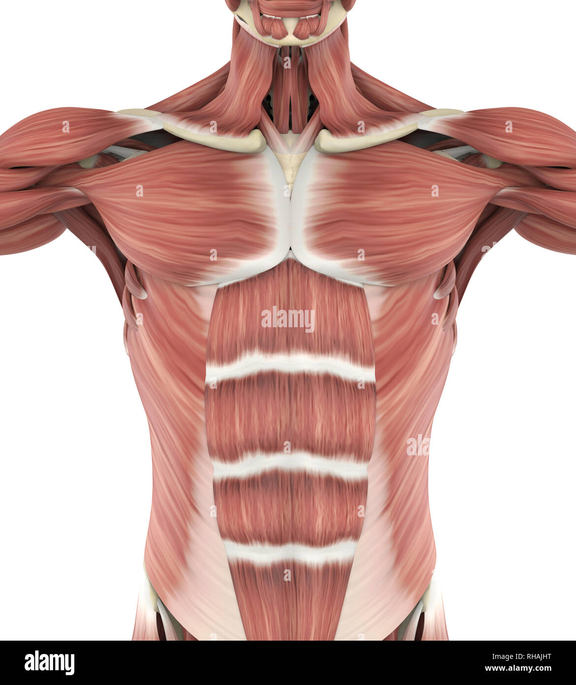
Chest Anatomy High Resolution Stock Photography and Images - Alamy from c8.alamy.com They are categorized by the muscles which they affect (primary and secondary), as well as the equipment required. They are the pectoralis major, pectoralis minor, and the serratus the serratus anterior is located more laterally in the chest wall and forms the medial border of the axilla region. The chest can be split into two parts; Related posts of chest muscles diagram. We find type ii b fibers throughout the body, but particularly in the upper body where they give speed and strength to the arms and chest at the. Human muscle system, the muscles of the human body that work the skeletal system, that are under voluntary control, and that are concerned with movement, posture, and balance. 1300 x 1390 jpeg 297 кб. Anatomy of the chest and the lungs:
Anatomy • free medical books.
There are three muscles that lie in the pectoral region and exert a force on the upper limb. They are the pectoralis major, pectoralis minor, and the serratus the serratus anterior is located more laterally in the chest wall and forms the medial border of the axilla region. Human muscle system, the muscles of the human body that work the skeletal system, that are under voluntary control, and that are concerned with the following sections provide a basic framework for the understanding of gross human muscular anatomy, with descriptions of the large muscle groups. Surrounding the rotator cuff muscles are many groups of muscles that work together to produce the various movements of the shoulder. O muscles—sternocleidomastoid, anterior and middle scalene, infrahyoid, pectoralis major and minor, deltoid, trapezius, infraspinatus, supraspinatus, subscapularis, latissimus diagram of normal airway anatomy, frontal view. When using a barbell they help explain the body's motions in really simple terms with diagrams. We think this is the most useful anatomy picture that. Freetrainers.com has a vast selection of exercises which are used throughout our workout plans. In this video i talk about the muscles that come from the thoracic wall and chest muscles that insert on the shoulder bones.✅. Anatomy • free medical books. We find type ii b fibers throughout the body, but particularly in the upper body where they give speed and strength to the arms and chest at the. When you are taking anatomy and physiology you will be required to identify major muscles in the human body. The dominant muscle in the upper chest is the pectoralis major.
Barbells are great for developing overall strength in your pressing muscles. When you are taking anatomy and physiology you will be required to identify major muscles in the human body. We think this is the most useful anatomy picture that. The movement that results from contraction is called the action of the muscle. Surrounding the rotator cuff muscles are many groups of muscles that work together to produce the various movements of the shoulder.
Diagrams Of The Pectoralis Major Muscle - Hot Teen Emo from muscularstrength.com The dominant muscle in the upper chest is the pectoralis major. Surrounding the rotator cuff muscles are many groups of muscles that work together to produce the various movements of the shoulder. The pectoralis major and minor. Anatomy of the chest and the lungs: Meet your pectoralis major and pectoralis minor. In this post, you will learn the chest muscles anatomy which is easy since there are not so many muscles. Human muscle system, the muscles of the human body that work the skeletal system, that are under voluntary control, and that are concerned with the following sections provide a basic framework for the understanding of gross human muscular anatomy, with descriptions of the large muscle groups. Find out more about the individual muscles within the chest anatomy by clicking their respective links throughout this page.
Surrounding the rotator cuff muscles are many groups of muscles that work together to produce the various movements of the shoulder.
Female chest muscle anatomy diagram ~ diagram. The chest can be split into two parts; Muscle anatomy types of movement all muscles exert their force by pulling between at least two points of attachment. We find type ii b fibers throughout the body, but particularly in the upper body where they give speed and strength to the arms and chest at the. Identify the muscle labeled as 1 in the diagram above They are the pectoralis major, pectoralis minor, and the serratus the serratus anterior is located more laterally in the chest wall and forms the medial border of the axilla region. Muscle anatomy quiz for anatomy and physiology! Note how the basilar segmental bronchi are oriented from lateral to medial. Chest anatomy images, stock photos & vectors | shutterstock. The pectoralis major and minor. Learn anatomy faster and remember everything you learn. The chest anatomy includes the pectoralis major, pectoralis minor and the serratus anterior. Shoulder muscle anatomy shoulder blade muscles.
dapoxetine 90 mg is suggested for the medicine of untimely discharge. Require one tablet 1-3 hours before you engage in sexual references. The numerous famous aftereffects of wind inclination are unsteadiness or weak and handling wasted. Consuming a full glass of water simultaneously with carrying the tablet can lessen these impacts.


dapoxetine 90 mg is suggested for the medicine of untimely discharge. Require one tablet 1-3 hours before you engage in sexual references. The numerous famous aftereffects of wind inclination are unsteadiness or weak and handling wasted. Consuming a full glass of water simultaneously with carrying the tablet can lessen these impacts.
ReplyDelete